AAV Purification Kits
For concentrating and purifying AAV vectors from cell lysate and cell culture media
For research use only and NOT intended for in vitro diagnostics.
AAV Purification Kits
For concentrating and purifying AAV vectors from cell lysate and cell culture media
Register today to receive an exclusive 15% off* on your first order.
Features and Benefits
- AAV Purification from any input – cell fraction or media fraction
- High AAV recovery, up to 90%
- No specialized equipment needed
- Purification from a variety of AAV serotypes (including AAV6 and AAV9)
- Yields highly active AAV for in vivo and in vitro experiments
- Purification is based on spin column chromatography that uses Norgen’s resin separation matrix
Recombinant adeno-associated virus (AAV) vectors are highly promising tools for both in vitro and in vivo gene transfer. Norgen’s AAV Purification Kits provide fast and simple procedures for concentrating and purifying AAV vectors from cell lysate and cell culture media. Purification is based on precipitation onto Norgen Biotek’s proprietary resin. Contaminating cellular debris is largely removed from the sample via a centrifugation step, while contaminating DNA and RNA is reduced using enzymatic digestion. AAV vector purified in this manner is highly active for use in in vitro and in vivo transduction experiments.
AAV Purification Kit
Norgen’s AAV Purification Kit contains sufficient materials for 15 preparations (33.5 mL per prep of supernatant (SN) or a total of 500 mL of supernatant input). Approximately 1 mL of cell pellet can be purified per prep, up to a maximum of 15 mL of cell pellet in total for the entire kit. Up to 33X sample concentration.
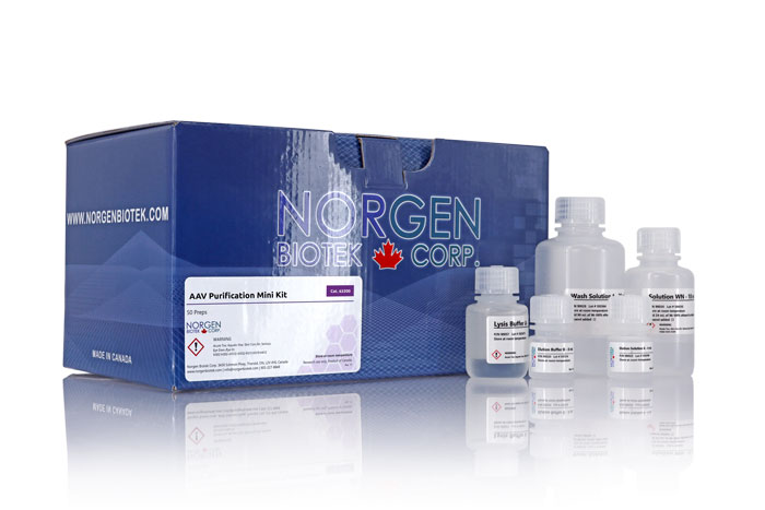
AAV Purification Mini Kit
Each spin column is able to concentrate and purify AAV from 0.5-8 mL of cell pellet, cell culture media, or cells and culture media mixed together. Up to 50X sample concentration. AAV vector purified in this manner is highly active for use in in vitro transduction experiments, and is eluted into a small volume (200 µL). Preparation time for 4 samples is 1.5 hours, with 45 minutes of hands-on time.
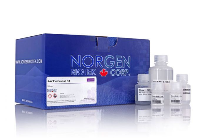
AAV Purification Midi Kit
Each spin column is able to concentrate and purify AAV from 8 mL up to 45 mL of input consisting of cell pellet, cell culture media, or cells and culture media mixed together. Up to 50X sample concentration. AAV vector purified in this manner is highly active for use in in vitro transduction experiments, and is eluted into a small volume (1 mL). The kit may be used to purify up to 8 x 25 mL or 4 x 45 mL of samples using the included columns. Preparation time for 4 samples is approximately 2 to 2.5 hours, with 1.5 hours of hands on time.
AAV Purification Maxi Kit (Slurry Format)
Each spin column is able to concentrate and purify AAV from 45 mL to 90 mL of input consisting of cell pellet, cell culture media, or cells and culture media mixed together. Up to 200X sample concentration. AAV vector purified in this manner is highly active for use in in vitro transduction experiments, and is eluted into a small volume (1-10 mL) using the optional concentration step. The kit may be used to purify up to 1 x 900 mL samples or 10 x 45-90 mL samples using the included columns. Preparation time for 1 x 900 mL sample is approximately 2.5 to 3.5 hours, with an optional concentration step requiring an additional 30 min.
Details
Supporting Data
Figure 1.In vitro transduction. HTX cells transduced with 50 µL eluted vector from the Norgen AAV Purification Kit after purification of cell culture supernatant containing AAV. Both AAV9 and a bovine AAV capsid (isolate AAV-Ca) were tested in vitro on HTX cells. The vector encoded an alkaline phosphatase reporter gene driven by the CAG promoter. Dark/purple staining represents cells that have been transduced by AAV bearing the alkaline phosphatase reporter gene.
Figure 2. In vivo testing of AAV vector purified using the Norgen AAV Purification Kit in the mouse lung. 2 x 10¹¹ ss genomes of an AAV9 vector encoding an alkaline phosphatase reporter gene was delivered via the intranasal route. 1 month post transduction, mouse tissues were harvested. Areas of transduction in the mouse lung are shown by the dark/purple staining where the alkaline phosphatase reporter gene was being expressed.
Figure 3. Transduction of HTX cells using Norgen’s AAV Purification Mini Kit. Microscopic view of HTX cells transduced with biologically active AAV vector after purification using Norgen's AAV Purification Mini Kit (dark purple represents alkaline phosphatase transgene expression).
Figure 4. Linear Scaling of Norgen's AAV Purification Mini Kit From 2.5 mL to 15 mL Input. Purification using Norgen's AAV Purification Mini Kit across different input volumes consisting of mixed cells and supernatant ranging from 2.5 mL to 15 mL. Increased input volumes resulted in no loss of vector as demonstrated by linear scaling across the different volumes.
Figure 5. Transduction of HTX cells with Norgen's AAV Purification Mini Kit with different input volumes of mixed cells and supernatant (0.5 mL. 2.5 mL, 10 mL and 15 mL). Microscopic view of HTX cells transduced with biologically active AAV vector after purification using Norgen's AAV Purification Mini Kit (dark purple represents alkaline phosphatase transgene expression).
Figure 6. Purification from Both Cell Pellet and Supernatant. Purification of AAV vector from both Cell Pellet and Cell Media/Supernatant fraction.
Figure 7. Linear Scaling of Norgen's AAV Purification Midi Kit From 2 mL to 50 mL Input. Purification using Norgen's AAV Purification Midi Kit across different input volumes of mixed cells and supernatant ranging from 2 mL to 50 mL. Increased input volumes resulted in no loss of vector as demonstrated by linear scaling across the different volumes.
Figure 8. Transduction of HTX cells with Norgen's AAV Purification Midi Kit with different input volumes (2 mL, 10 mL and 50 mL) of mixed cells and supernatant. Microscopic view of HTX cells transduced with biologically active AAV vector after purification using Norgen's AAV Purification Midi Kit (dark purple represents alkaline phosphatase transgene expression).
Figure 9. Cellular Transduction of HTX cells with 50 µL of AAV purified using Norgen’s AAV Purification Maxi Slurry Kit. Dark purple staining indicates expression of a human placental alkaline phosphatase reporter gene from AAV transduced cells.
Figure 10. Supernatant from cells transfected with AAV production plasmids was purified using the Norgen AAV Purification Maxi Slurry Kit. Three different volumes were tested: 40, 60 and 80 mL, demonstrating scalable purification of AAV vector over increasing volumes.
|
Kit Specifications
|
|
| Resin Binding Capacity (total per kit) | At least 5 x 10¹⁰ AAV particles as determined by qPCR |
| AAV Vector Serotype | AAV6, AAV9 and others |
| Input Type | Cells, media |
| Input Volume (AAV supernatant) | 1 - 33.5 mL SN per prep (500 mL SN in total) |
| Input Volume (AAV cell pellet) | 1 mL cell pellet per prep (15 mL in total) |
| Time to Complete Purifications | 2.5 to 4.5 hours with 1 hour hands on time |
| In vivo transduction | Yes |
Storage Conditions and Product Stability
HL-SAN Nuclease should be stored at -20°C upon arrival. Elution Buffer O should be stored tightly capped at 4°C upon arrival. All other solutions should be kept tightly sealed and stored at room temperature. Once opened, the solutions should be stored at 4°C. This kit is stable for 2 years after the date of shipment.
| Component | Cat. 66100 (15 preps) | Cat. 63200 (20 preps) | Cat. 63300 (4-8 preps) | Cat. 63250 (1-10 preps) |
|---|---|---|---|---|
| Lysis Buffer S | 5.5 mL | 5.5 mL | 5.5 mL | 20 mL |
| DNAse I | - | 2 x 25 uL | 2 x 25 uL | 210 μL |
| RNAse A | - | 60 μL | 60 μL | 240 μL |
| HL-SAN Nuclease | 102 μL | - | - | - |
| Binding Buffer A | 20 mL | 4 mL | 4 mL | 2 x 8 mL |
| Purification Solution C | 60 mL | - | - | - |
| Purification Solution D | 130 mL | - | - | - |
| Wash Solution C | 2 x 130 mL | 60 mL | 60 mL | 3 x 60 mL |
| Slurry E | 12.5 mL | - | - | 2 x 14.5 mL |
| Elution Buffer O | 66 mL | 8.5 mL | 8.5 mL | 66 mL |
| Protein Neutralizer | 4 mL | 4 mL | 4 mL | 4 mL |
| Spin Columns | - | 20 | - | - |
| Mini Spin Columns | - | 20 | - | - |
| Midi Spin Columns (grey contents) with Collection Tubes | - | - | 8 | 10 |
| Midi Spin Columns (white contents) with Collection Tubes | - | - | 8 | - |
| Maxi Spin Columns (grey contents) with Collection Tubes | - | - | - | 10 |
| Maxi Spin Columns (white contents) with Collection Tubes | - | - | - | 10 |
| Collection Tubes | - | 40 | - | - |
| Elution tubes (1.7 mL) | 50 | 20 | - | - |
| Midi Elution tubes (15 mL) | - | - | 8 | 10 |
| Maxi Elution tubes (50 mL) | - | - | - | 10 |
| Product Insert | 1 | 1 | 1 | 1 |
Documentation
FAQs
Mini
AAV sample does not flow through the column due to one or more of the following:
- Centrifugation speed was too low.
Check the centrifuge to ensure that it is capable of generating 1,500 x g. Sufficient centrifugal force is required to move the liquid phase through the resin. - Inadequate spin time.
Spin an additional two minutes to ensure that the liquid is able to flow completely through the column. - Cell debris obstructing column.
Ensure that no cell debris is inadvertently applied to the column following lysate centrifugation. Care should be taken to ensure that only the supernatant is applied to the column. Filtration of supernatant through a 0.2 µm or 0.45 µm filter may decrease clogging of the column. The sample can also be divided among several columns.
Poor AAV recovery could be due to one or more of the following:
- Incorrect pH adjustment of AAV sample for binding.
A pH level of 3.5-3.8 works best for binding AAV to the resin. Ensure that the sample is adjusted to approximately this value with Binding Buffer A prior to sample loading. In phenol red containing media, this will turn the media a bright yellow color. - Initial titer of the sample applied to the column was too low.
A sufficient amount of AAV particles is required in the starting sample to ensure success in downstream applications. It may be required to increase the volume of the starting sample or increase the AAV vector titer of the starting sample by optimizing transfection conditions and/or vector constructs. - Elution Buffer O stored improperly.
Ensure that the Elution Buffer O is aliquoted into 1.5 mL microfuge tubes after opening, taking care to minimize the amount of air left in the tubes (1 mL to 1.5 mL aliquots). Store tightly capped at 4°C.
If a pellet is observed following elution, the input may have been too high for a single column. Spin down elution containing the pellet, and harvest the supernatant, as the AAV vector should mostly be present in the supernatant. The pellet can be re-processed using a fresh column with the cell pellet protocol to harvest the remaining AAV present in the pellet.
Midi
AAV sample does not flow through the column due to one or more of the following:
- Centrifugation speed was too low. Check the centrifuge to ensure that it is capable of generating 1,500 x g. Sufficient centrifugal force is required to move the liquid phase through the resin.
- Inadequate spin time. Spin an additional two minutes to ensure that the liquid is able to flow completely through the column.
- Cell debris obstructing column. Ensure that no cell debris is inadvertently applied to the column following lysate centrifugation. Care should be taken to ensure that only the supernatant is applied to the column. Filtration of the supernatant through a 0.2 µm or 0.45 µm filter may decrease clogging of the column. The sample can also be divided among several columns.
Poor AAV recovery could be due to one or more of the following:
- Incorrect pH adjustment of AAV sample for binding. A pH level of 3.5-3.8 works best for binding AAV to the resin. Ensure that the sample is adjusted to approximately this value with Binding Buffer A prior to sample loading. In phenol red-containing media, this will turn the media a bright yellow color.
- Initial titer of the sample applied to the column was too low. A sufficient amount of AAV particles is required in the starting sample to ensure success in downstream applications. It may be required to increase the volume of the starting sample or increase the AAV vector titer of the starting sample by optimizing transfection conditions and/or vector constructs.
- Elution Buffer O stored improperly. Ensure that the Elution Buffer O is aliquoted into 1.5 mL microfuge tubes after opening, taking care to minimize the amount of air left in the tubes (1 mL to 1.5 mL aliquots). Store tightly capped at 4°C.
If a pellet is observed following elution, the input may have been too high for a single column. Spin down the elution containing the pellet, and harvest the supernatant, as the AAV vector should mostly be present in the supernatant. The pellet can be re-processed using a fresh column with the cell pellet protocol to harvest the remaining AAV present in the pellet.