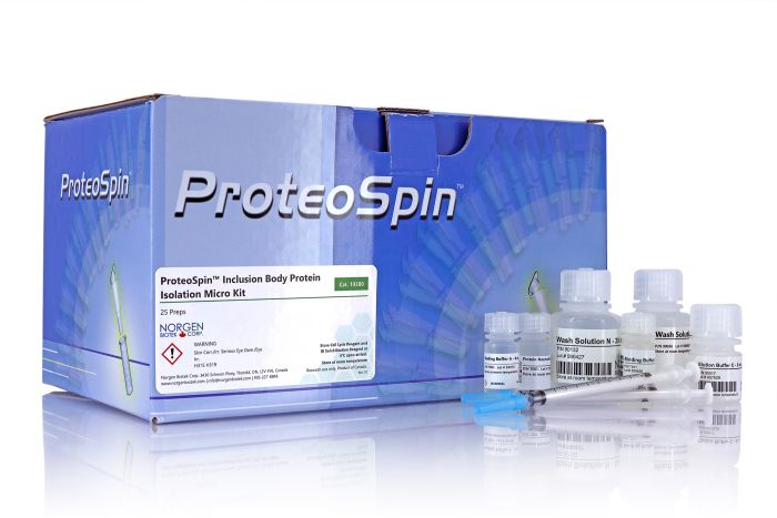
ProteoSpin™ Inclusion Body Protein Isolation Kits
For the rapid isolation of inclusion body proteins

For research use only and NOT intended for in vitro diagnostics.
ProteoSpin™ Inclusion Body Protein Isolation Kits
For the rapid isolation of inclusion body proteins
Register today to receive an exclusive 15% off* on your first order.
Features and Benefits
- All-in-one solution for inclusion body protein isolation and purification
- Fast and convenient spin column protocol
- Complete kit with Cell Lysis Reagent, Inclusion Body Solubilization Reagent, buffers and spin columns to purify proteins
- Purification is based on spin column chromatography that uses Norgen’s resin separation matrix
These kits provide everything required to isolate and purify inclusion body proteins from induced bacterial cultures. First a proprietary Cell Lysis Reagent is used to selectively lyse the cells and release inclusion bodies in their solid form. Next, inclusion bodies are dissolved and their contents released using the provided IB Solubilization Reagent. Inclusion body proteins are then further purified using spin columns for rapid and convenient buffer exchange and desalting. This kit provides a convenient way to screen recombinants prior to scaling up.
ProteoSpin™ Inclusion Body Protein Isolation Micro Kit
The process is efficient and streamlined and can process up to 12 samples in only 60 minutes. Each spin column is able to recover up to 50 µg of acidic or basic proteins. Purified recombinant proteins are then ready for SDS-PAGE, 2D gels, Western blots, Mass Spectrometry analysis, and other applications.
ProteoSpin™ Inclusion Body Protein Isolation Maxi Kit
The procedure is efficient and streamlined and can process up to 4 samples in approximately 2 hours. Each spin column is able to recover up to 12 mg of acidic or basic proteins from 100 mL of induced bacterial culture. Purified recombinant proteins are then ready for SDS-PAGE, 2D gels, Western blots, Mass Spectrometry analysis, and other applications.
About Inclusion Bodies
Bacteria are widely used for the expression of different proteins. However, 70-80% of the proteins expressed in bacteria by recombinant techniques are typically contained in insoluble inclusion bodies (i.e., protein aggregates). The protein of interest found in these subcellular structures is often inactive, due to incorrect folding. The production rate of recombinant proteins stored in inclusion bodies is invariably higher than those synthesized as soluble proteins. The reason behind this is thought to be the resistance of insoluble proteins to proteolysis by cellular enzymes. In addition, separation of insoluble recombinant proteins in inclusion bodies is considerably easier than that of soluble proteins. These factors have been the major influences favoring scale-up of high-value proteins using bacterial fermentation for example. Procedures for the purification of the expressed proteins from inclusion bodies are often labour-intensive, time-consuming and not cost-effective. This kit provides the essential reagents for cell disruption, inclusion body solubilization and purification using spin column chromatography - all optimized to work together thereby simplifying the process and saving a tremendous amount of time and cost.
Details
Supporting Data
Figure 1. Isolation of Acidic Recombinant Protein. Small (1.5 mL) cultures of BL21 (DE3) pLysS bacteria expressing a 50 kDa chimeric protein (acidic) were pelleted. Cells were lysed and inclusion bodies separated and dissolved using the ProteoSpinTM Inclusion Body Protein Isolation Micro Kit. The resulting protein was bound to the ProteoSpinTM column, washed and eluted in 50 µL of the provided elution buffer. The eluted protein samples were analyzed in 12.5% polyacrylamide gels, which were run for 45 minutes at 200 V/6.5 cm. The protein bands were made visible by staining with Coomassie Blue R-250.
Figure 2. Isolation of Basic Recombinant Protein (RNase A). Small (1.5 mL) cultures of BL21 (DE3) pLysS bacteria expressing RNaseA (basic) were pelleted. Cells were lysed and inclusion bodies separated and dissolved using the ProteoSpinTM Inclusion Body Protein Isolation Micro Kit. The resulting protein was bound to the ProteoSpinTM column, washed and eluted in 50 µL of the provided elution buffer. The eluted protein samples were analyzed in 12.5% polyacrylamide gels, which were run for 45 minutes at 200 V/6.5 cm. The protein bands were made visible by staining with Coomassie Blue R-250.
Figure 3: Efficient Isolation of Inclusion Body Proteins. Following gene expression, inclusion bodies were extracted, solubilized and proteins were purified using Norgen's Inclusion Body Protein Isolation Kits. Briefly, cells were pelleted and lysed using the provided Cell Lysis Reagent. The inclusion bodies were separated via centrifugation and washing and dissolved using the Inclusion Body Solubilization Reagent. Recovered proteins were analyzed on 12% SDS-PAGE and stained with Coomassie Brilliant Blue R-250. Numbers represent kDa sizes of protein bands. As can be seen, proteins of a wide range of masses are efficiently purified. Lane numbers represent size of protein purified in kDa.
Figure 4: No Loss of Proteins when Isolating a Basic Protein. 100 mL of induced bacterial culture expressing a recombinant 30 kD BASIC protein were pelleted and processed using the ProteoSpinTM Inclusion Body Protein Isolation Maxi Kit. Briefly, pelleted cells were lysed, inclusion bodies were separated and subsequently dissolved using the provided Inclusion Body. Solubilization Reagent. Fractions of input, flowthrough, wash and elution were loaded on a 12.5% acrylamide gel. Lane 1 is the input, Lane 2 is the flowthrough from the input, Lane 3 is the wash, Lane 4 is the first elution and Lane 5 is the second elution. As can be seen, proteins are not lost in the flowthrough or wash. Recombinant proteins were efficiently bound to the column and eluted.
Figure 5: No Loss of Proteins when Isolating An Acidic Protein. 100 mL of induced bacterial culture expressing a recombinant 70 kD ACIDIC protein were pelleted and processed using the ProteoSpinTM Inclusion Body Protein Isolation Maxi Kit. Briefly, pelleted cells were lysed, inclusion bodies were separate and subsequently dissolved using the provided Inclusion Body Solubilization Reagent. Fractions of input, flowthrough, wash and elution were loaded on a 12.5% acrylamide gel. Lane 1 is the input, Lane 2 is the flowthrough from the input, Lane 3 is the wash, Lane 4 is the first elution and Lane 5 is the second elution. As can be seen, proteins are not lost in the flowthrough or wash. Recombinant proteins were efficiently bound to the column and eluted.
|
Kit Specifications
|
|
| Maximum Culture Volume |
1.5 mL
|
| Yield from 1.5 mL |
Up to 50 μg
|
| Minimum Elution Volume |
30 μL
|
| Time to Process 12 Samples |
60 minutes
|
Storage Conditions
The Cell Lysis Reagent and IB Solubilization Reagent should be stored at 4°C upon receipt of this kit. All other solutions should be kept tightly sealed and stored at room temperature. This kit is stable for 2 years after the date of shipment. Once opened, the solutions should be stored at 4°C when not in use except for the pH Binding Buffers (Acidic and Basic). Some precipitation will occur with 4°C storage. This precipitation should be dissolved with slight heating to room temperature before using.
| Component | Cat. 10300 (Micro - 25 preps) | Cat. 17700 (Maxi - 4 preps) |
|---|---|---|
| Wash Solution C | 30 mL | 130 mL |
| Wash Solution N | 30 mL | 130 mL |
| Binding BUffer A | 4 mL | 20 mL |
| Binding Buffer N | 4 mL | 20 mL |
| Elution Buffer C | 8 mL | 2 x 30 mL |
| Protein Neutralizer | 4 mL | 4 mL |
| Cell Lysis Reagent | 15 mL | 110 mL |
| IB Solubilization Reagent | 2 mL | 50 mL |
| Syringes, 1cc, slip tip | 25 | - |
| Needles (Bev, 20G x 1 inch) | 25 | - |
| Syringes, 10 mL, Luer-Lok™ Tip | - | 4 |
| Needles (18G x 1.5 inch) | - | 4 |
| Micro Spin Columns | 25 | - |
| Maxi Spin Columns (filled with SiC) inserted into 50 mL collection tubes | - | 4 |
| Collection Tubes | 25 | - |
| Elution Tubes (1.7 mL) | 25 | - |
| Elution Tubes (50 mL) | - | 4 |
| Product Insert | 1 | 1 |
Documentation
FAQs
Micro, Maxi
No inclusion body pellet may be observed due to:
- Improper induction of gene expression.
Consult the manufacturer’s expression system literature.
- Gene product does not produce inclusion bodies.
Reassess the expression cassette.
Inefficient cell lysis may be occurring due to the following:
- Kit solutions were improperly stored.
Keep the lysis and solubilization reagent at 4℃ at all times when not in use. The two binding buffers should be kept at room temperature.
- Freeze/thaw step was not performed.
The freeze/thaw step is known to increase lysis efficiency. Repeat the protocol using the recommended freeze/thaw conditions.
- Lysozyme may be required to increase lysis efficiency.
Add lysozyme to concentrations recommended by the supplier.
- Mechanical disruption of cells was inefficient.
Increase the number of passages through the needle and syringe.
Column clogging can result from the following:
- Centrifugation speed was too low.
Check the centrifuge to ensure that it is capable of generating the required speed for a particular step. Sufficient centrifugal force is required to move the liquid phase through the resin.
- Inadequate spin time.
Spin an additional 2 minutes to ensure that the liquid is able to flow completely through the column.
- Protein solution is too viscous.
Dilute the protein solution and adjust the pH accordingly. Highly viscous materials due to high protein concentration can retard the flow rate.
- Cellular debris is present in the protein solution.
Filter the sample with a 0.45 µM filter or spin down insoluble materials and transfer the liquid portion to the column. Solid, insoluble materials can cause severe clogging problems.
- Protein solution is not completely dissolved.
Repeatedly vortex and pipette to try and dissolve inclusion bodies. If needed, incubate on a rocker at room temperature to aid in solubilization. Solid, insoluble materials can cause severe clogging problems.
This may be because of the liberation of nucleic acids following lysis. Increase the degree of mechanical disruption by passing bacteria / lysis reagent through the needle at least five more times. Alternatively, add an appropriate amount of DNase I.
The eluted protein forms a precipitate when:
- Protein is too concentrated.
Vortex and repeatedly pipette to try and create a homogeneous solution. If needed, heat slightly to return the protein into solution.
- pH of eluted protein is close to the protein’s pI.
Check the pH to verify it is the same as the pI of the protein. Add additional Protein Neutralizer to bring the pH away from the pI of the protein (~1 pH lower or higher).
Poor protein recovery could be due to one or more of the following:
- Incorrect procedure was used.
Ensure that the acidic protocol was used for an acidic protein, and the basic protocol for a basic protein. It is known that when basic proteins are bound using the acidic protocol, elution is inefficient because the basic proteins are bound tightly.
- Incorrect pH adjustment of sample.
Ensure that the pH of the sample is 4.5 for acidic proteins and 7.0 for basic proteins.
- Protein may have precipitated prior to loading onto the column.
If the pH of the protein sample is the same as the pI of your protein, precipitation may occur. In this case, adjust the pH of your sample to at least one pH unit lower than the pI of your protein.
The eluted protein may be degraded due to one or more of the following:
- Eluted protein solution was not neutralized.
Add an appropriate amount of Protein Neutralizer to the eluted protein. Some proteins are sensitive to high pH, such as the Elution Buffer C at pH 12.5.
- Eluted protein solution was not neutralized quickly enough.
If eluted protein is not used immediately, degradation will occur. We strongly suggest adding Protein Neutralizer to lower the pH.
- Proteases may be present.
Use protease inhibitors during all steps of sample preparation.
- Bacterial contamination of the protein solution.
Prepare the protein samples with 0.015% sodium azide.
Too many gel bands can be caused by inefficient cell lysis which may be due to one or more of the following:
Kit solutions were improperly stored. Keep the lysis and solubilization reagent at 4°C at all times, when not in use. The two binding buffers are kept at room temperature.
Freeze / thaw step was not performed. The freeze / thaw step is known to increase lysis efficiency. Repeat the protocol using the recommended freeze / thaw conditions.
Lysozyme may be required to increase lysis efficiency. Add lysozyme to concentrations recommended by supplier.
Mechanical disruption of cells was inefficient. Increase the number of passages through the needle and syringe.
Citations
Maxi (17700)
| Title | CP40 from Corynebacterium pseudotuberculosis is an endo-β-N-acetylglucosaminidase |
| Journal | BMC Microbiology. 2016. |
| Authors | Shadnezhad A, Naegeli A, Collin M. |
| Title | Proteome of Human Calcium Kidney Stones. |
| Journal | Urology. 2010. |
| Authors | Canales BK, Anderson L, Higgins L, Ensrud-Bowlin K, Roberts KP, Wu B, Kim IW, Monga M. |
