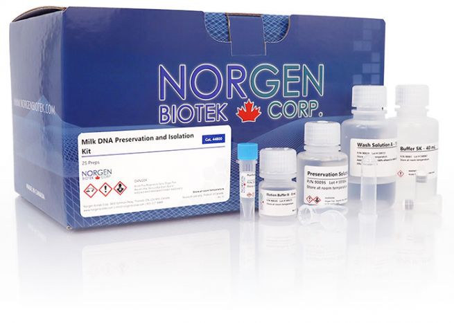
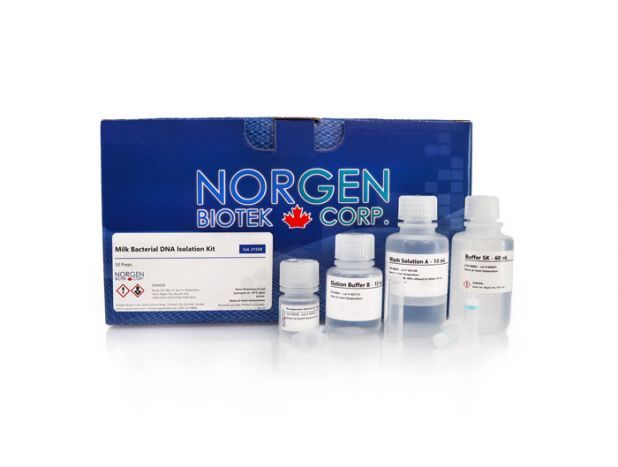
Milk Bacterial DNA Isolation Kit
For research use only and NOT intended for in vitro diagnostics.

For research use only and NOT intended for in vitro diagnostics.