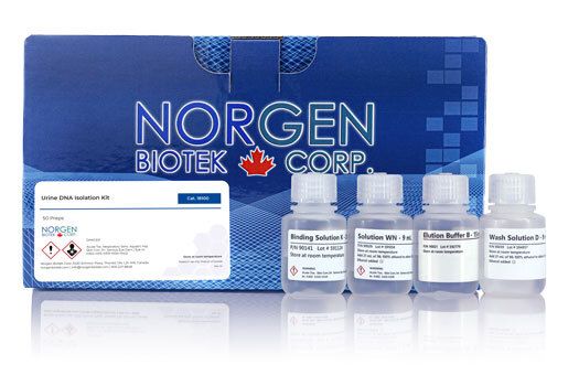
Urine DNA Isolation Kits

For research use only and NOT intended for in vitro diagnostics.
CE-IVDD marked diagnostic slurry format available here
Urine DNA Isolation Kits
Register today to receive an exclusive 15% off* on your first order.
Supporting Data
Figure 1. A Typical Agarose Gel Showing Total Urinary DNA Isolated from 1.5 mL of Urine using Norgen's Urine DNA Isolation Kit. Total urinary DNA was isolated from two different 1.5 mL urine samples. The samples were processed according to the bind, wash and elute procedure provided in the kit, and the DNA was eluted into 2 separate elution volumes. The proteins were first eluted into 100 µL of Elution Buffer (E1), followed by a second elution using 75 µL of Elution Buffer (E2). The purified urine DNA was then run on a 1.2% agarose gel, and each ane contains one tenth from each elution (i.e. E1: 10 µL out of 100 µL were loaded on the gel, E2: 7.5 µL out of 75 µL were loaded on the gel ). Lane M is 10 µL of Norgen’s FastRunner DNA Ladder. Both the large and small urine DNA species can be seen on the gel.
Figure 2. Isolation and Detection of DNA from 1.75 mL Urine Samples. Total genomic DNA was isolated from two different 1.75 mL urine samples using Norgen’s Urine DNA Isolation Kit. The bind, wash and elute procedure was performed, and the purified DNA was eluted into two separate elutions of 100 µL (E1) and 75 µL (E2). The isolated DNA was then subjected to quantitative PCR using human 5S gene primers to detect the genomic DNA using the iQ SYBR Green Supermix (BioRad, #170-8882). Five microliters from each elution was used as a template in a 20 µL qPCR reaction. The red line in the PCR baseline graph above corresponds to the first elution from the DNA isolated from the first urine sample, the green line corresponds to the second elution from the DNA isolated from first urine sample, the blue line corresponds to the first elution from the DNA isolated from the second urine sample, whereas the orange line corresponds to the second elution from the DNA isolated from the second urine sample. The yellow line corresponds to the No Template Control.
Figure 3. Circulating DNA Isolated from Urine can be used as the Template in PCR Reactions.Total urinary DNA was isolated from three different 1.5 mL urine samples using Norgen's Urine DNA Isolation Kit. The bind, wash and elute procedure was performed, and the purified DNA was eluted into two separate elutions of 100 µL (E1) and 75 µL (E2). Five microliters of each elution was then used as a template in a PCR reaction to amplify the K-ras gene. Lanes A-C contain the expected 157 bp product, and correspond to the first elution from each sample. Lane D is the positive control of 293 HEK DNA and shows the expected 157 bp product, while Lane E is the negative control. Lane M is Norgen's FastRunner DNA Ladder.
Figure 4. Typical Agarose Gel Showing Total Urinary DNA Isolated from Different Urine Volumes using Norgen's Urine DNA Isolation Kit (Slurry Format). Total urinary DNA was isolated from 3 mL, 6 mL, 12 mL and 24 mL of urine. Total urinary DNA was isolated from each urine sample according to the isolation protocol that is optimized for different sample volumes. The isolated DNA was eluted into two separate elutions (E1 and E2). The purified urine DNA was then loaded onto a 1.5% agarose gel. Each lane shows one tenth from each elution. It can be seen that the first elution contains most of urinary DNA whereas the second elution contains the rest of the urinary DNA isolated. Lane M is 10 µL of Norgen's FastRunner DNA Ladder.
Figure 5. Isolation and Detection of DNA from Different Volumes of Urine. Total genomic DNA was isolated from 3, 6, 12 and 24 mL urine samples using Norgen's Urine DNA Isolation Kit (Slurry Format). The urinary DNA was isolated from each urine sample according to the provided protocol, and the DNA was eluted into two separate elutions (E1 and E2). The isolated DNA was then subjected to quantitative PCR using human 5S gene primers to detect the genomic DNA. To test the absence of PCR inhibitors usually accompanying DNA isolated from urine, an increasing amount from each elution (1, 3, 6 and 9 µL) was used as a template in the PCR reaction. Urinary genomic DNA was successfully detected from all the different urine volumes with no sign of PCR inhibition, even when 9 µL of urine DNA was used as the template.
Figure 6. Highly Sensitive Isolation of Viral DNA from 25 mL of Urine. Herpes Simplex Virus - 1 (HSV-1) sequences were cloned into a plasmid, and 25 mL urine samples were then spiked with decreasing amounts of the plasmid (106, 105, 104, 103, 102 and 101). Total DNA was then isolated using Norgen’s Urine DNA Isolation Kit (Slurry Format). Ten microlitres of each 100 µL elution was then used as the template in a 20 µL PCR reaction using HSV-1-specific primers and Norgen's 2X PCR Master Mix (Cat #28007). Each PCR reaction was then run on a 1.5% agarose gel for visual analysis. The HSV-1 amplification product is 275 bp. As few as 250 HSV-1 viral copies could be isolated and detected from 25 mL of urine.
Figure 7. Typical Agarose Gel Showing Total Urinary DNA Isolated from Different Urine Volumes using Norgen's Urine DNA Isolation Maxi Kit (Slurry Format). Total urinary DNA was isolated from 20 mL, 40 mL, 60 mL and 80 mL of urine. Total urinary DNA was isolated from each urine sample according to the isolation protocol that is optimized for different sample volumes. The isolated DNA was eluted, and was then loaded onto a 1.8% agarose gel. Each lane shows one tenth from each elution. It can be seen that urine DNA is increasing linearly with the urine input volume. It should also be noted that the circulating DNA started to appear in the form of ladder with the increase of the urine sample input. Lane M is 10 µL of Norgen's FastRunner DNA Ladder.
Figure 8. Linearity of Ct Values and DNA Yields When Scaling Up Urine Input. Total urinary DNA was isolated from 20 mL, 40 mL, 60 mL and 80 mL of urine. Total urinary DNA was isolated from each urine sample according to the isolation protocol that is optimized for different sample volumes. The isolated DNA was then subjected to quantitative PCR using human 5S gene primers to detect the genomic DNA. Urinary genomic DNA was successfully detected from all the different urine volumes with no sign of PCR inhibition and in a linear relationship to the total urine DNA yield (Ct values decrease with increasing the sample urine input)
|
Kit Specifications
|
|
|
Volume of Urine Processed
|
1.75 mL
|
|
Average Yield*
|
Up to 50 ng
|
|
Size of DNA Purified
|
Large (> 1 kb)
and small (150-250 bp) |
| Time to Complete 10 Purifications |
30 minutes hands-on time
(plus a 1 hour incubation) |
* Yield will vary depending on the type of sample processed
Storage Conditions and Product Stability
All buffers should be kept tightly sealed and stored at room temperature. This kit is stable for 2 years after the date of shipment. The kit contains a ready-to-use Proteinase K, which is dissolved in a specially prepared storage buffer. The buffered Proteinase K is stable for up to 2 years after the date of shipment when stored at room temperature.
| Component | Cat. 18100 (50 preps) | Cat. 48800 (50 preps) | Cat. 50100 (50 preps) |
|---|---|---|---|
| Binding Solution K | 15 mL | - | - |
| Slurry B1 | - | 18 mL | 18 mL |
| Proteinase K in Storage Buffer | 2 mL | - | - |
| Pronase K in Storage Buffer | 2 mL | - | - |
| Soluton WN | 9 mL | - | - |
| Binding Buffer A | - | - | 50 mL |
| Lysis Buffer A | - | 30 mL | 30 mL |
| Wash Solution A | - | 38 mL | 38 mL |
| Wash Solution B | 30 mL | - | - |
| Wash Solution D | 9 mL | - | - |
| Binding Solution K | 15 mL | - | - |
| Elution Buffer B | 15 mL | 15 mL | 15 mL |
| Micro Spin Columns | 50 | - | - |
| Mini Filter Spin Columns | - | 50 | 50 |
| Collection Tubes | 50 | 50 | 50 |
| Elution Tubes (1.7 mL) | 100 | 100 | 100 |
| Product Insert | 1 | 1 | 1 |
Documentation
A Novel Method To Capture Methylated Human DNA From Urine Implications For Prostate Cancer Screening - Ascb 2008
The Urinary Genomic and Proteomic Profiling of Hepatocellular Carcinoma Patients Infected With Hepatitis C Virus - ASM 2008
Sensitivity of DNA Extraction Methods from Different Bodily Fluids for Human Identification
FAQs
Spin Column, Slurry, Maxi Slurry
RPM= √RCF/(1.118x10-5)(r)
Where RCF = required gravitational acceleration (relative centrifugal force in units of g); r = radius of the rotor in cm; and RPM = the number of revolutions per minute required to achieve the necessary g-force.
We recommend the following steps to prepare frozen urine for isolation:
- Gently warm the sample to room temperature or 37°C for 5 min.
- DO NOT perform a centrifugation step - this will eliminate the precipitated proteins leading to loss of protein-bound cf-NA or exosomes.
- Proceed with the protocol.
We recommend the use of Norgen’s Urine Preservative when collecting urine samples. Norgen’s Urine Preservative is designed for the preservation of nucleic acids and proteins in fresh urine samples at ambient temperatures, therefore no protein precipitation will occur and the purified nucleic acids will be of a higher quality.
